
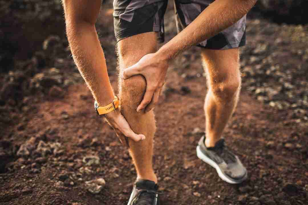
Arthritis after an ACL tear — the short answer
An ACL-injured knee carries a higher lifetime risk of osteoarthritis, and ACL reconstruction does not bring that risk back to baseline. In the classic cohort (Lohmander et al., Arthritis & Rheumatism 2004), about half of female soccer players had radiographic knee arthritis 12 years after injury, reconstructed or not. Meniscus preservation, quad strength, body weight, and early staging change the odds. Individual risk varies.
- Risk is elevated but not destiny — many ACL patients never need a replacement
- The meniscus is the biggest surgical lever: repair beats removal for decades of cartilage health
- Quad strength and body weight are the levers you control after surgery
- Early arthritis is the treatable kind — a standing X-ray and an exam are all it takes to stage it
Reconstruction doesn't reset the clock
That's not a reason to skip surgery. Reconstruction restores stability, protects the meniscus from the repeated giving-way episodes that shred it, and gets athletes back to the activities they love. But it's a reason to think of an ACL tear as a knee-health event with a long tail — one that deserves attention years after the graft has healed. Most patients hear a lot about the operation and the rehab year, and almost nothing about decade two. This article is about decade two. This type of joint wear after injury is called post-traumatic arthritis, and understanding why it happens points directly at what you can do to slow it down.
Why a stable, "fixed" knee can still wear out
The damage starts at the moment of injury. When the ACL tears, the joint surfaces slam together hard enough to bruise the bone — MRI shows this bone bruise in most acute tears — and that impact injures cartilage cells in ways no operation can undo. Reconstruction rebuilds the ligament; it cannot rebuild the cartilage that was hit.
Then there's what tore alongside the ACL. Meniscus and cartilage injuries travel with ACL tears, and losing meniscus tissue concentrates load on a smaller area of cartilage for the rest of the knee's life. Finally, even a well-reconstructed knee doesn't move exactly like the original — subtle changes in rotation and load distribution can shift wear onto cartilage regions that weren't built for it. Add the quad weakness that lingers when rehab is cut short, and the joint absorbs more shock with less muscular protection. None of these factors is destiny, but together they explain why the risk stays elevated.
What actually lowers your risk
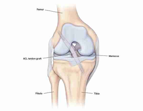
You can't change the injury, but several of the biggest risk factors are in your hands — or your surgeon's. The single most important surgical factor is the meniscus: knees that keep their meniscus do meaningfully better over decades than knees that lose it, which is why Dr. Morton repairs tears whenever the tissue allows rather than removing them. If you're facing that decision, read about meniscus repair surgery before agreeing to a meniscectomy.
After surgery, the levers are yours — and two matter more than the rest. A strong quadriceps acts as the knee's shock absorber, unloading cartilage with every step, which is why finishing rehab completely pays dividends for decades. And body weight multiplies across the knee joint with every stride, so even modest weight loss meaningfully reduces the load your cartilage carries through a lifetime of steps.
- Preserve the meniscus — choose repair over removal whenever the tear allows
- Finish rehab completely; a strong quadriceps shields cartilage from impact for years
- Keep body weight in a healthy range — every extra pound multiplies across the knee with each step
- Stay active with joint-friendly cardio: swimming, cycling, paddling, hiking on moderate grades
- Take giving-way episodes seriously — recurrent instability accelerates cartilage damage
- Don't ignore new swelling, aching, or stiffness in a previously quiet knee
Monitoring: what surveillance of an ACL-injured knee looks like
You don't need an MRI every year — you need a low-effort habit of paying attention. For most patients, sensible monitoring means a periodic check-in with a knee specialist if symptoms change: new aching after activity, swelling that shows up after workouts, morning stiffness, or a slow loss of the activities the knee used to tolerate. A standing X-ray is a simple, inexpensive way to track joint-space changes over time, and comparing films across visits answers most questions. Patients who lost meniscus tissue at the original surgery, or who notice the knee drifting bow-legged or knock-kneed, deserve a lower threshold for checking in.
Early detection matters because early arthritis is the treatable kind. Activity adjustments, strength work, weight management, and injections all work better on a joint that still has cartilage to protect. Waiting until the knee is bone-on-bone skips every rung of that ladder. If your ACL injury was years ago and the knee is starting to talk to you, that's exactly the right moment for a consultation — not a reason to wait until it shouts.
If arthritis develops: you have options at every stage
A diagnosis of post-traumatic arthritis is the start of a management plan, not a countdown to a replacement. Early on, most patients do well with strength training, activity modification, weight management, anti-inflammatories, and injection therapies — including orthobiologic options like platelet-rich plasma (PRP), framed honestly (see PRP for knee arthritis). A focal cartilage lesion in an otherwise preservable knee may be a candidate for cartilage restoration. There's a full menu of alternatives to knee replacement worth exhausting first, especially in younger patients.
When arthritis is confined to one compartment — common after ACL injury, where the inner compartment often wears first — a partial knee replacement can resurface just the damaged area and preserve everything else. And if the whole joint eventually wears out, modern total knee replacement is a durable, well-proven operation. The point isn't that surgery is inevitable; for many ACL patients it never happens. The point is that at every stage, from a twinge at forty to a worn joint at sixty, there's a next move — and the earlier you engage, the more moves you have. Age alone doesn't disqualify anyone from any rung of the ladder; the state of the cartilage, your symptoms, and your goals drive the plan.

Get your knee staged — your next step
Get your knee staged. An exam and standing X-rays tell you where you are — book a consultation or call (808) 439-6201; Dr. Morton sees patients in Honolulu (Ala Moana), Kunia, Hilo, and Kona. Bring any prior imaging and your operative report if you have one; managed-care and Medicaid/Quest plans may need a referral from your primary care physician, which the office checks before your visit. Neighbor island patients can start by telehealth with local X-rays when an in-person visit isn't practical.
- Joint-preserving options are likely if: pain is activity-related, X-rays show joint space remaining, and the damage is focal or confined to one compartment
- It's time to discuss replacement if: the knee is bone-on-bone on standing X-rays, pain limits daily life despite strength work and injections, and you've exhausted the alternatives
Medically reviewed by Paul Norio Morton, MD, FAAOS, FAAHKS. Last reviewed: . This article is for education only and isn’t a substitute for an exam and personalized advice — schedule a consultation to discuss your situation.