
What is avascular necrosis of the hip?
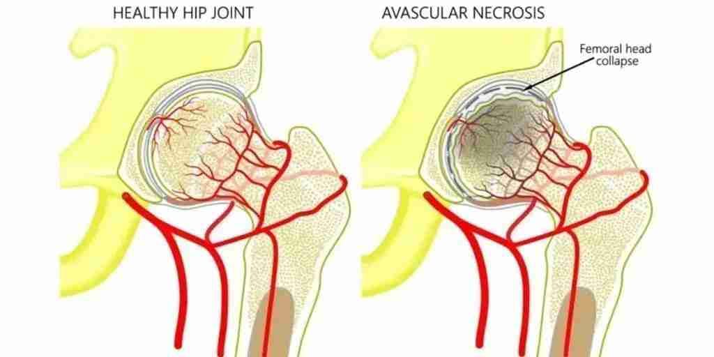
The hip is a ball-and-socket joint where two bones meet: the head of the thigh bone (the femur) sits inside a cup-shaped part of the pelvis called the acetabulum. Avascular necrosis — also called osteonecrosis — is the death of bone in the femoral head, the ball of the joint, caused by an interruption of its blood supply.
Bone is living tissue. Without circulation, the affected segment weakens, and the round head can eventually fracture and collapse like a failing archway. Once the ball loses its shape, the cartilage above it fails too, and the result is rapidly progressive arthritis, pain, and difficulty walking.
What makes AVN different from ordinary wear-and-tear arthritis is who it affects: patients are frequently in their 30s, 40s, and 50s — working, raising families, and far from the age most people associate with hip disease. Pain usually starts in the groin, worsens with weight-bearing, and can appear in both hips, since the underlying causes act on the whole body rather than one joint. As the disease advances, stiffness and a limp follow, and pain begins to show up at rest and at night. Many patients first see their primary care physician for the pain, but most ultimately reach an orthopedic surgeon for definitive treatment.
What causes AVN?
AVN rarely has a single trigger. Most researchers believe it develops when several factors act at once to create a kind of "perfect storm" that starves the femoral head of blood. It can affect children or adults, and the causes fall into a few broad groups.
The two most common culprits
- Corticosteroids (steroids). Prolonged or high-dose glucocorticoids — prescribed for many medical conditions — can impair blood flow to the femoral head and lead to bone death.
- Heavy alcohol use. Excess alcohol is thought to promote AVN through changes in stress hormones, an increase in circulating fat, and sluggish blood flow through the small vessels that feed the bone.
Medical conditions and other risks
- Lupus and the presence of antiphospholipid antibodies
- Sickle cell disease and Gaucher disease
- Decompression sickness ("the bends") in divers
- Clotting disorders
- Acute lymphoblastic leukemia and kidney transplantation
- Trauma — a fall, a car crash, a hip fracture or dislocation can physically tear the blood vessels feeding the femoral head
- In children, a congenital condition called Legg-Calvé-Perthes disease
In a substantial share of patients no cause is ever found; this is called idiopathic AVN. It matters less than people fear — treatment is driven by the stage of the disease, not its origin. What does matter is acting on the diagnosis: AVN tends to progress, and the difference between an early hip and a collapsed one is the difference between preserving your joint and replacing it.
How AVN is diagnosed and staged
Pinning down the stage of AVN is the single most important step, because it decides whether the joint can be saved. Dr. Morton works through a structured evaluation.
History and examination
- Symptom assessment. When did the pain start, and is it getting worse? A pattern of steadily increasing groin pain is an important clue.
- Medical history review. A careful look for risk factors — previous hip injury, corticosteroid use, heavy alcohol use, and the conditions associated with AVN.
- Physical examination. Testing the hip's range of motion to find the positions that reproduce discomfort and to gauge how the joint is moving.
Imaging and testing
- X-rays give a baseline view of the bone's density and shape, but they can look completely normal in early disease.
- MRI is the most sensitive test for pre-collapse AVN. It reveals damage to bone and marrow long before an X-ray does, and it shows the size and location of the lesion — details that drive treatment.
- Blood tests don't stage the disease directly, but they help identify underlying causes and contributing factors.
With that information, the disease is placed in an early, middle, or late stage, each reflecting a different level of bone and joint damage. Early stages lend themselves to joint-preserving treatment; advanced stages point toward replacement.
Stages: why timing changes everything
Beneath the staging systems lies one essential question: has the femoral head collapsed?
In early, pre-collapse disease, the bone is injured but the ball is still round. This is the window in which joint-preserving surgery can work — and it is why an MRI is ordered whenever AVN is suspected, even when the X-ray looks normal.
Once the head collapses, a crescent-shaped fracture line appears, the ball flattens, and the cartilage is destroyed. From that point, no procedure can restore the native joint, and treatment shifts to replacement. Because that window closes silently, unexplained groin pain in a younger adult — especially with steroid exposure or heavy alcohol use — deserves prompt evaluation. Schedule a consultation early; the stage at diagnosis largely determines your options.
Treatment options: from managing risk factors to surgery
The first step in any AVN treatment plan is to find the cause and stop or manage the offending agent — for example, reducing steroid exposure where medically possible or addressing heavy alcohol use — while optimizing every other factor that impedes blood flow, including quitting smoking.
Early on, some patients try non-operative management: reducing weight-bearing with crutches and using medications to control pain and preserve bone density. It can ease symptoms, but studies have consistently shown that non-operative care does not reliably halt the disease's progression toward collapse. For that reason, many patients move on to surgery, which divides into two families of procedures:
- Joint-preserving surgery — aimed at saving the patient's own femoral head, best suited to earlier, pre-collapse disease.
- Joint replacement — the reliable answer once the head has collapsed.
Hip preservation: core decompression with BMAC
For pre-collapse AVN, the workhorse joint-preserving procedure is core decompression. Through a small incision, one or more channels are drilled into the dead segment of the femoral head. This relieves the painful pressure inside the bone and creates pathways for new blood vessels and healing bone to grow into the lesion. Many surgeons then pack the channel with a bone graft to fill the areas where bone has died.
The research on core decompression is genuinely mixed: many patients get significant pain relief, but the best, most durable results are concentrated in those treated early, before any collapse, with smaller lesions. Results are less reliable for large lesions or once collapse has begun.
That is why Dr. Morton frequently augments core decompression with bone marrow aspirate concentrate (BMAC) — marrow drawn from your pelvis and spun down to concentrate mesenchymal stem cells and growth factors, then delivered into the decompressed channel. Adding these cells has been shown to improve outcomes over decompression alone: patients tend to report greater reductions in hip pain and better joint function, and the biology appears to slow the progression of AVN — lowering the risk that the femoral head collapses and, with it, the likelihood of eventually needing a total hip replacement. It is part of a broader orthobiologics approach to keeping young joints out of the operating room.

The stem-cell (BMAC) procedure, step by step
Combining core decompression with concentrated stem cells is a precise, image-guided operation designed to regenerate bone using the patient's own tissue. Here is what it involves.
1. Bone marrow collection
The procedure begins by drawing bone marrow from the iliac crest — the rim of the pelvis you can feel at your waist — through a needle. The marrow is placed in tubes and spun in a centrifuge, which concentrates the stem cells and growth factors and separates them from the rest of the marrow.
2. Planning the decompression
Using advanced imaging such as a CT scan, a three-dimensional model of the hip is built to map the exact regions of the femoral head affected by AVN, so the dead bone can be targeted precisely.
3. The surgical process
Through small incisions near the hip — each roughly a centimeter long — a guide is placed into the femoral head. A navigated drill follows that guide to remove the necrotic bone. The concentrated stem cells are then injected into those areas, along with a bone graft to support healing.
4. Recovery and aftercare
The operation is typically outpatient, so most patients go home the same day; those with significant pain may stay briefly. Afterward, weight-bearing is usually protected — many patients stay toe-touch weight-bearing with crutches or a walker for about two weeks while the bone begins to heal, then progress back to most normal activities. Medications are prescribed both to manage pain and to reduce the risk of blood clots, and regular follow-up visits track how the bone is healing.
Risks
Complications are uncommon but possible, and they include infection, bleeding, and soreness at the marrow-collection site. More serious problems — bone fracture, blood clots, or cartilage damage — are rare. Dr. Morton will review the specific risks and benefits for your situation before surgery.
How stem cells help rebuild the bone
Stem cells are unspecialized cells with the ability to develop into the body's many specialized cell types. Broadly, they fall into two groups: embryonic stem cells, which can become almost any cell type, and adult (somatic) stem cells, which live in tissues such as bone marrow and drive the body's everyday repair.
AVN treatment uses adult stem cells, and the logic is straightforward:
- Collection — the cells come from your own bone marrow, which improves compatibility and eliminates any risk of rejection.
- Transplantation — they are placed directly into the damaged areas of the femoral head.
- Regeneration — once in place, they help clear away dead bone and encourage the growth of healthy new bone tissue.
By harnessing the body's own healing machinery, this targeted regeneration aims to address the damage AVN has already done while supporting long-term recovery of mobility and comfort.
When the head has collapsed: hip replacement
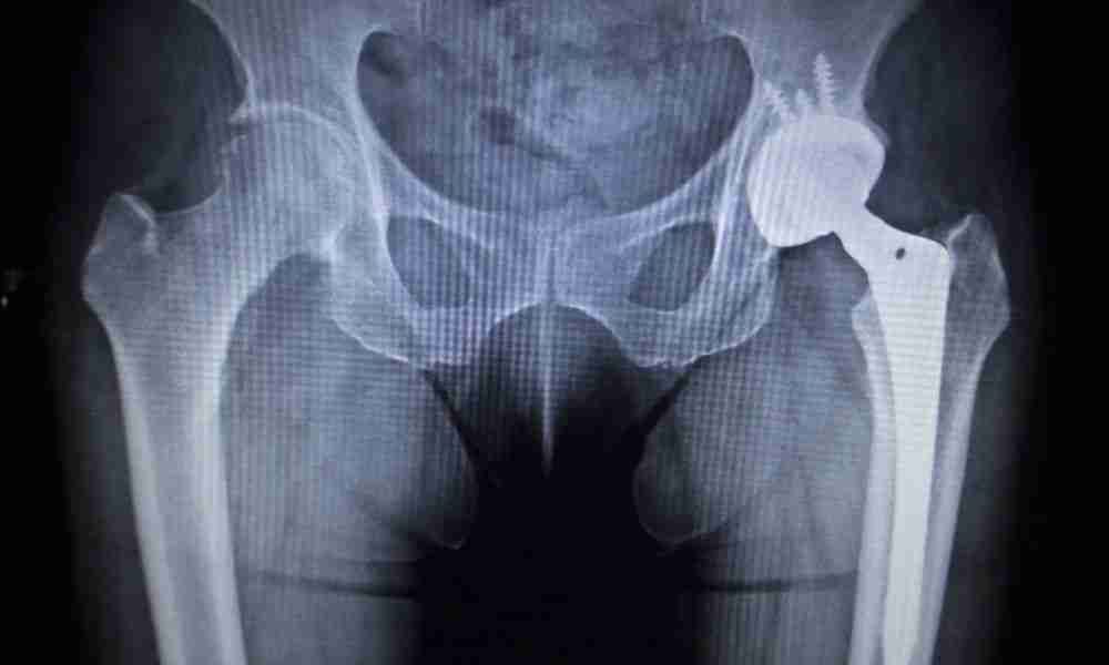
Once the femoral head has collapsed, preservation procedures rarely succeed, and total hip replacement becomes the reliable answer — it removes the dead bone entirely and resurfaces the socket, resolving the pain at its source. Total hip replacement is one of the most successful operations in medicine, with a low revision rate, which is why some surgeons favor it over joint-preserving surgery once the disease is advanced.
Many AVN patients hesitate because of their age. It's a fair concern, and it's addressed head-on in Am I too young for a hip replacement? — the short version is that modern cementless implants and improved bearings have made replacement a sound option for younger, active patients when the joint is already destroyed. Dr. Morton performs these through the muscle-sparing anterior approach, usually with no hip precautions afterward, with precise component positioning and cementless implants that let your bone grow directly into the prosthesis.
Because AVN is frequently bilateral, both hips are evaluated and some patients ultimately need both replaced. Dr. Morton prefers to stage those surgeries — one hip, then the other once you've recovered — rather than replacing both on the same day, which is safer and makes rehabilitation more manageable. Living with a collapsed hip to "save" the replacement for later often means years of limping, stiffness, and lost fitness that make eventual surgery harder.
What affects the outlook (prognosis)?
No two cases of AVN are identical, and the outlook depends on several factors that Dr. Morton weighs when he builds a plan:
- Underlying cause. Whether AVN stems from trauma, steroids, or a systemic disease can influence how it responds to treatment.
- Stage at diagnosis. Early detection, before significant bone damage, generally leads to better outcomes — the single biggest lever you and your surgeon have.
- Location and size of the lesion. Damage to critical, weight-bearing areas of the femoral head can be harder to treat.
- Age and general health. Younger, healthier, more active patients often heal more efficiently.
- Treatment approach. The specific procedure chosen shapes the long-term result.
The best way to protect your prognosis is to seek evaluation promptly, follow your treatment plan, and keep the controllable risk factors — smoking, alcohol, and overall health — in check.
Medically reviewed by Paul N. Morton, MD, FAAOS, FAAHKS — board-certified orthopedic surgeon, fellowship-trained in adult hip & knee reconstruction and orthopedic trauma.
This page is for education only and isn’t a substitute for an exam and personalized advice — schedule a consultation to discuss your situation.